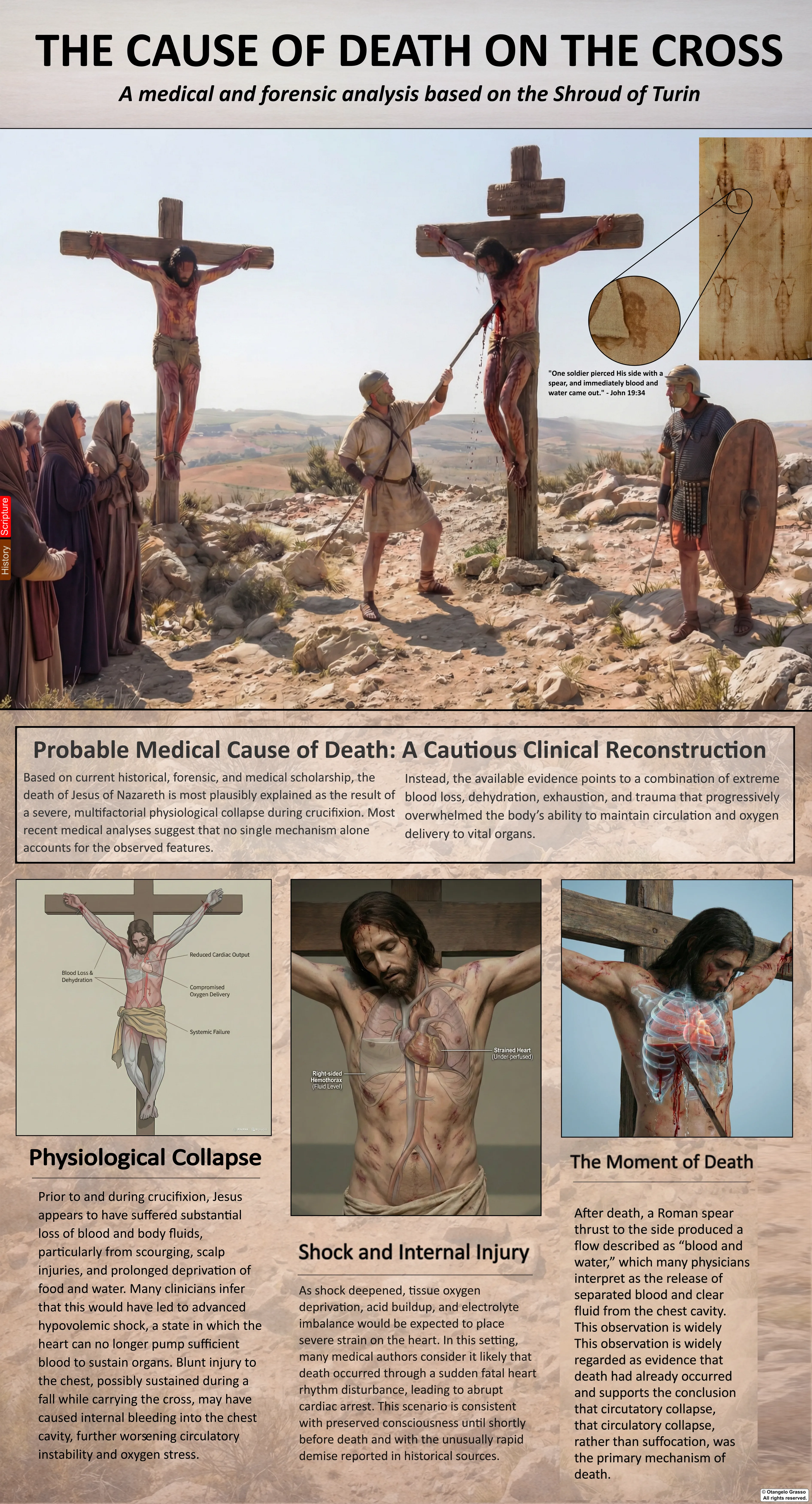
The accounts agree that Jesus died on the cross. They do not give us a clinical record of how his body failed. Between that historical event and a modern medical explanation lies the work of reconstruction.
Mark describes Pilate’s surprise that Jesus was already dead, followed by an inquiry to the centurion before permission was given to release the body. John describes soldiers finding him dead, leaving his legs unbroken, and piercing his side. These are consequential details. They establish the sequence within the narratives, but they do not supply a pulse tracing, a measured blood loss, or an examination of the heart and lungs. The task is to ask which physiological processes could account for death under the reported conditions, and what additional information the Shroud might contribute if its image and wound-associated stains derive from a crucified body.1
This chapter brings together the medical discussions in The Shroud of Turin: A Forensic Summary of the Evidence, From Forensics to Faith, and The Image That Should Not Exist; the original books of Pierre Barbet and Frederick Zugibe; the relevant papers in the supplied collection; and the principal published debates about crucifixion physiology. The result is a critical narrative review, not a retrospective death certificate. Established physiology, historical testimony, experimental findings, and Shroud-specific interpretation are identified separately throughout.2, 3, 4, 5, 6, 7
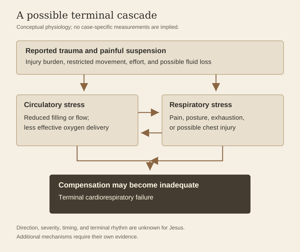
A combined failure of circulation and respiration is a medically plausible framework for Jesus’s death after severe trauma and crucifixion. Blood and fluid loss, pain, exhaustion, posture, and possible chest injury could have interacted. The evidence does not establish their relative contributions, demonstrate a unique terminal mechanism, or allow measured values to be assigned to Jesus’s blood gases, electrolytes, or cardiac rhythm.
1. What the historical record actually supplies
The canonical Gospels describe an execution carried out by Roman authority under Pontius Pilate. Their Passion narratives report beating, mockery, scourging, crucifixion, death, and burial, with differences in sequence and detail. Mark 15:25 places crucifixion at the third hour, while John 19:14 places the sentencing scene at about the sixth hour. Mark and Matthew associate the death with the ninth hour. Translating these expressions into a single synchronized modern timetable requires historical and literary judgments; the texts do not authorize a six-hour physiological chart with exact measurements assigned to each hour.1
The frequently used expression “six-hour anomaly” should therefore be handled carefully. A death within the same day can be compared with reports of longer survival, and Mark explicitly presents Pilate as surprised. Nevertheless, there is no representative Roman dataset giving a normal survival time, standard wound burden, or standard position on a cross. A rapid death does not select one cause from several possibilities. It might reflect the injuries before crucifixion, a particular arrangement of the body, individual vulnerability, an additional complication, or interacting processes.1, 4, 8
Josephus supplies a useful comparison in Life 420–421. He recounts recognizing three acquaintances among crucified captives and obtaining their removal and medical care. Two subsequently died; one recovered. The passage demonstrates that removal while alive could sometimes be followed by survival. It does not tell us how long those individuals had been suspended, whether their injuries matched Jesus’s, or whether their treatment resembles an unaided recovery in a tomb.9
The Gospel details also have limits that reconstructions sometimes cross unnoticed. John does not specify the side pierced, the intercostal space, the length of the wound tract, or the organ struck. Mark does not describe a centurion feeling for a carotid pulse. The sources do not narrate an electrocardiographic arrest, ventricular fibrillation, or a visibly separated sequence of dark blood followed by clear serum. These may enter a medical model, but they must remain additions to the record rather than details presented as eyewitness testimony.1
2. Cause, mechanism, and circumstance of death
Three questions need different answers. The circumstance is an execution by crucifixion. The injuries and conditions are those reported in the texts or proposed from the image: scourging, puncture wounds, loss of blood and fluid, prolonged painful suspension, and possibly thoracic injury. A terminal mechanism describes the physiological failure through which death occurred: for example, critically inadequate circulation or ineffective gas exchange. “The heart stopped” names an endpoint shared by many causes; it does not identify the injury or disease responsible.2, 3, 8
A modern examiner ordinarily combines an external examination with internal dissection, medical history, scene investigation, histology, toxicology, and sometimes imaging. For Jesus there is no body available for that examination. For the Shroud there is an image, a textile, and limited sampled material whose provenance and interpretation must themselves be assessed. Robert Bucklin’s “autopsy” is valuable as an expert reading of the depicted injuries, but it is a simulated examination of an image. It cannot establish internal lesions with the certainty available at an actual autopsy.10
It is also important to separate Jesus’s death from the identity of the Shroud figure. A medical interpretation of crucifixion does not depend on accepting the cloth’s authenticity. Conversely, an anatomically plausible crucifixion pattern on the cloth does not alone identify the person or date the textile. The two inquiries can inform one another, but using the Gospel narrative to identify every mark and then using those same identifications to “independently” confirm the narrative would be circular.
3. The injuries before suspension
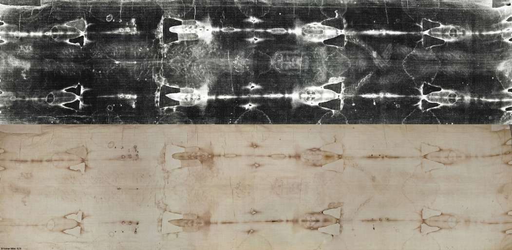
Crucifixion did not begin with the nails. The reported scourging and beating matter because a person arriving at the cross after significant trauma has less reserve than an uninjured volunteer. Skin and deeper soft-tissue wounds can bleed externally, retain blood internally, and provoke inflammation. Damaged tissue can swell as fluid leaves the circulation. Pain, exertion, reduced intake, and sweating can increase the overall burden. These processes can interact without any single visible wound being sufficient by itself to explain death.2, 3, 11, 12
The Shroud is often interpreted as showing many paired or grouped scourge marks across the torso and limbs. Counts vary with photograph quality, enhancement, overlap, and the definition of a separate mark. A count of visible marks cannot be multiplied by an assumed number of millilitres per mark to obtain a reliable blood-loss total. The image does not reveal wound depth, the calibre of injured vessels, the time each wound bled, or how much fluid was lost before the cloth was applied.4, 5
Scalp injuries add pain and can bleed because the scalp is well supplied with vessels. That does not establish that thorn punctures alone caused massive haemorrhage. Similarly, a proposed fall, chest contusion, fractured rib, or lung laceration must not be silently promoted to a documented injury. Such lesions would be clinically important if present; neither the Gospel descriptions nor the Shroud image demonstrate their presence with the specificity of radiography or dissection.3, 4
Luke’s description of sweat becoming like drops of blood has sometimes been diagnosed as haematidrosis. The wording, textual history, and relation between comparison and literal bleeding require discussion before a rare diagnosis is assigned. Even if literal bloody sweating is entertained, it cannot supply a measured contribution to hypovolaemia. The larger medical question is the cumulative burden of trauma, not whether every devotional detail can be converted into a named disorder.1, 3
4. How blood loss can become circulatory failure
Shock means that the circulation cannot adequately meet tissue requirements. In haemorrhagic shock, loss of circulating blood reduces the volume returning to the heart. With less filling, the heart may eject less blood with each beat. A faster pulse and constriction of peripheral vessels can initially protect blood flow to vital organs, but these compensations have limits. A person can remain conscious during part of that process; consciousness does not establish normal circulation.3, 12
The problem is oxygen delivery, which depends on blood flow as well as the blood’s oxygen content. Blood loss can impair both: there is less circulating volume and, after haemorrhage and fluid redistribution, less haemoglobin available to carry oxygen. Thus a person may be in dangerous shock even if the lungs are still moving air. Conversely, a reassuring oxygen saturation measured in a healthy suspension volunteer would not test the effects of severe blood loss in an injured victim.
Thirst in John’s narrative is compatible with bodily distress, including fluid deficit, but it is not a laboratory test for shock. Simon of Cyrene’s carrying of the cross in the Synoptic accounts can fit exhaustion or weakness, yet does not establish a particular haemorrhage class. Describing Jesus as having lost 30–40 percent of his blood or having a heart rate of 150 beats per minute gives a reconstruction clinical precision it has not earned.1, 4, 11
The case for a circulatory contribution is therefore mechanistic rather than numerical: the reported injuries and prolonged painful execution create credible pathways to inadequate perfusion. The amount and rate of loss remain unknown. Zugibe develops this argument at length, treating traumatic and hypovolaemic shock as central to the fatal process. His reconstruction is medically coherent, while its unobserved lesions and quantities remain hypotheses about this particular death.3
5. Pain, exhaustion, and upright immobility
Pain is not an alternative to physiology; it changes physiology. Sympathetic activation can increase heart rate and the demand placed on the myocardium. Muscular effort consumes oxygen. Pain in an injured chest can lead to shallow breathing to avoid rib movement, a response called splinting. Restricted movement and prolonged upright posture can also alter venous return. These effects make a combined model more plausible than an account that isolates blood loss, posture, or respiratory effort from the rest of the injury burden.3, 12
Neither “traumatic shock” nor “neurogenic shock” should be used as a vague synonym for extreme suffering. Neurogenic shock has a specific clinical meaning associated with loss of sympathetic vascular tone, commonly after spinal cord injury. No such injury is established here. Reflex circulatory events associated with pain are possible, but their occurrence in Jesus cannot be inferred merely because the pain would have been severe.
Modern harness-suspension research offers a limited comparison. Rauch and colleagues investigated circulatory responses in suspended volunteers and identified a potential reflex contribution to presyncope. Harness geometry, pressure on the body, freedom of the limbs, and support differ from a cross. These experiments show why immobility deserves attention, not that modern suspension syndrome is a confirmed diagnosis for Jesus.13
The same distinction applies to exhaustion. It is reasonable to consider exhaustion after trauma and hours of execution. It is not reasonable to infer a precise remaining muscle reserve, a fixed interval between attempts to move, or the second at which the victim could no longer support himself. A useful reconstruction explains how reserve could fail without pretending to have watched the failure unfold.
6. Barbet’s model: expiration becomes a struggle
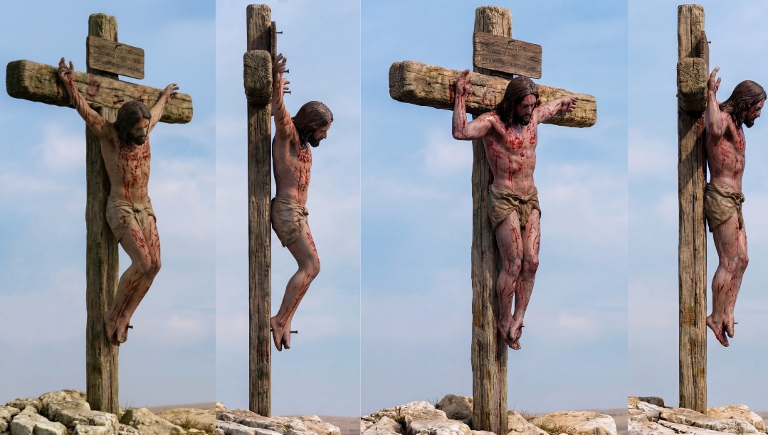
Pierre Barbet’s A Doctor at Calvary gave the best-known account of crucifixion as an asphyxial process. In his model, traction through the arms and sustained contraction of respiratory muscles fix the chest toward an inspiratory position. Expiration becomes difficult. The victim must reduce the pull on the arms by raising the body against the feet, obtaining temporary respiratory relief before fatigue causes renewed sagging. Barbet interpreted aspects of the Shroud’s chest, abdomen, and wrist flows within this model.2
The proposal is more specific than saying crucifixion makes breathing painful. It asserts a repeated mechanical obstruction to effective ventilation, a required movement to overcome it, and a progressive inability to sustain that movement. Each part needs evidence. A cloth image showing a chest contour cannot itself measure ventilation, and two apparent blood-flow directions cannot establish the frequency or purpose of movements on the cross.
Barbet also drew on descriptions of suspension torture in which the hands were raised high and the feet unsupported. Such testimony concerns real suffering, but the mechanical arrangement matters. Near-vertical arm suspension is not interchangeable with an arrangement in which the arms extend more laterally, the feet bear some load, or the person can change posture. One cannot infer a universal respiratory mechanism without accounting for those differences.2, 14
Even the supplied edition of Barbet’s book contains dissent. In Appendix II, P. J. Smith argues for shock from exhaustion, pain, and blood loss rather than accepting Barbet’s asphyxia explanation. The controversy did not begin with recent skeptical objections: it was present within the early medical discussion itself.2
7. Zugibe’s experiments and the challenge to asphyxia
Frederick Zugibe tested aspects of crucifixion posture using volunteers secured with restraints rather than nails. His studies examined breathing, oxygenation, discomfort, and the feasibility of movements proposed by Barbet. He reported maintained or increased oxygenation in the conditions tested, increasing abdominal breathing, and no need to repeatedly raise the body to breathe. He also disputed the interpretation of the wrist bifurcation as a record of two breathing positions.3
This evidence is important because it moves beyond a verbal model to an observed response. In the tested configuration, suspension did not automatically produce the respiratory failure predicted by a strong version of the classical account. A model that says the posture alone must rapidly prevent adequate breathing is weakened if people can breathe adequately in that posture.
The limits are equally important. Healthy volunteers in short, supervised experiments had not been scourged, dehydrated, or nailed. Restraints distribute load differently from penetrating injuries; the subjects could report symptoms and be released. Consequently, those experiments cannot establish that breathing remained adequate in a severely injured person over hours. They challenge an obligatory posture-driven asphyxia mechanism; they do not exclude respiratory contribution from pain, lung injury, exhaustion, or later shock.3
Zugibe’s own explanation still includes substantial physiological distress. His disagreement with Barbet concerns the dominant mechanism and the proposed necessity of lifting for each effective breath. It should not be rewritten as a claim that crucifixion is mechanically harmless or that an injured chest cannot compromise respiration.
8. Why the modern reviews disagree
Edwards, Gabel, and Hosmer’s influential 1986 JAMA article proposed hypovolaemic shock combined with exhaustion asphyxia. It remains a major reference because it joins the reported trauma to respiratory and cardiovascular consequences. Its detailed anatomical reconstruction, however, is a reconstruction from historical material rather than a contemporaneous medical examination.11
Maslen and Mitchell’s 2006 review compared competing medical explanations and emphasized weaknesses in the underlying evidence and the use of historical sources. Their conclusion was that the available record does not establish exactly how crucifixion victims died, with posture and circumstances likely affecting the mechanism. That is a methodological constraint, not a claim that death by crucifixion is medically inexplicable.8
Habermas, Kopel, and Shaw’s 2021 survey characterized asphyxia or asphyxia-dominant explanations as the leading position among the views they assembled. McGovern, Kaminskas, and Fernandes subsequently challenged the evidential basis of the suffocation model, especially the transfer of conclusions from forms of hanging torture to more familiar cross configurations. The latter paper appeared online in 2022 and in the 2023 journal volume.14, 15
These publications answer different questions: what physicians have proposed, how often a view appears in a selected literature, and how directly its mechanism is supported. Repetition cannot turn a hypothesis into an observation. Nor does a later critique, simply by being later, settle every uncertainty. The responsible position is to compare the evidence for each component and acknowledge that published medical disagreement continues.
9. A terminal cascade rather than an invented clinical chart
A combined model begins with the possibility that injuries have reduced circulating volume and increased oxygen demand. Pain and posture may make breathing less efficient. Reduced tissue perfusion can increase metabolic stress; reduced ventilation, if present, adds a separate gas-exchange problem. These processes can reinforce one another: a poorly perfused respiratory muscle may tire more easily, while impaired oxygenation increases the burden on an already stressed circulation.3, 4, 12
The distinction between hypoxaemia and inadequate tissue oxygen delivery is useful here. Hypoxaemia refers to reduced oxygen in arterial blood. A person can also suffer inadequate delivery because the heart is circulating too little blood or because haemoglobin has been lost. These pathways can coexist, but none should be assumed merely from an image of a distressed body.
In severe shock, metabolic acidosis can develop as oxygen supply, cellular metabolism, and the clearance of metabolic products become impaired. If ventilation is also inadequate, carbon dioxide retention may produce respiratory acidosis. The interaction can further impair cardiovascular performance. This is an explanatory mechanism drawn from trauma physiology, not a measurement of Jesus’s pH.3, 4, 16
Similarly, a severely stressed heart could develop a lethal rhythm disturbance or fail to maintain effective circulation without ventricular fibrillation. No electrocardiogram exists to distinguish ventricular fibrillation, profound bradycardia, asystole, or pulseless electrical activity. A final cry followed by death does not identify the rhythm. The endpoint is observable in the narratives; the electrical sequence is not.
10. Coagulation: an additional possibility, not a diagnosis
Joseph Bergeron proposed traumatic shock complicated by trauma-induced coagulopathy as a possible contributor, and potentially a major mechanism, in Jesus’s death. The attraction of this proposal is that severe injury can impair the body’s ability to control bleeding; a person’s deterioration need not be explained solely by the initial volume lost. Bergeron explicitly recognizes that the exact mechanism or combination cannot be proven.12
Modern trauma research describes interacting changes in the vascular lining, platelets, clotting factors, and fibrinolysis. Depending on circumstances and timing, the response can favour bleeding or thrombosis. It is not simply a process in which all blood loses the ability to clot. Visible external clots could therefore coexist with systemic derangement, while visible clots alone do not demonstrate that derangement.16
For Jesus, there are no coagulation studies, platelet counts, or tissue samples sufficient to diagnose a systemic disorder. For the Shroud, fluid-looking transfers cannot establish that the person suffered trauma-induced coagulopathy before death. Postmortem blood behaviour and the later transfer process introduce separate explanations. The proposal belongs in the differential assessment, with its modern physiological basis distinguished from the absent case-specific tests.7, 17
11. What cannot be reconstructed from the chemistry
Some narratives assign a particular blood pH, potassium concentration, respiratory rate, or oxygen saturation to successive stages of crucifixion. Others describe a predictable progression from rhabdomyolysis to hyperkalaemia and a named fatal arrhythmia. Muscle injury, impaired renal function, and acid–base disturbances can matter in trauma. The existence of those pathways does not demonstrate their magnitude or even their occurrence in this historical case.4
It is especially misleading to make a very short period without food into a necessary major ketoacidotic crisis in an otherwise unspecified adult. Nor is potassium change in complex trauma adequately captured by an automatic one-for-one exchange of hydrogen ions for potassium. These details require measurements and clinical context that are unavailable.
The same restraint applies to temperature. The modern trauma triad of coagulopathy, acidosis, and hypothermia is clinically important, but hypothermia cannot be assigned to Jesus from an assumed weather report. A physiological model should include only the conditions for which there is a justified historical or experimental basis, and should identify everything else as conditional.16
12. Cardiac rupture and haemopericardium
A separate family of proposals locates the decisive event in the heart. Haemopericardium means blood within the sac surrounding the heart. If accumulation raises pressure enough to prevent adequate cardiac filling, tamponade can follow. A rupture of the heart wall is one possible route to that emergency. This mechanism differs from a heart that stops because the whole circulation has failed after blood loss or respiratory impairment.2, 3
Luigi Malantrucco developed a post-infarction haemopericardium interpretation discussed in the author’s Forensic Summary. It tries to explain a relatively rapid death and the later appearance of blood and clear fluid within one anatomical account. It requires an antecedent myocardial lesion, a rupture, an appropriate accumulation of fluid, and an outlet reached by the spear. The supplied book’s discussion documents the proposal; it does not supply an independently examined infarct or a specimen of the pericardium.4, 18
A right-sided wound does not make a cardiac pathway impossible. The heart occupies the mediastinum rather than lying entirely in the left chest, and the right atrium contributes to its rightward contour. Trajectory, entry position, body orientation, blade length, and depth all matter. A model can reach the heart from the right. Conversely, a stain on a flat textile cannot determine that route or establish which chamber was entered. An exact tract length would require assumptions about anatomy and wound position that the cloth cannot resolve.3
Even demonstrated blood in the pericardium would not automatically establish infarction as its cause. Penetrating injury, other cardiac lesions, and different sources of bleeding would need consideration. The pre-existing terminal lesion must be separated from damage caused by the spear after death.
13. Stress cardiomyopathy and sudden cardiac death
Recent literature has reconsidered a cardiac explanation in terms of stress cardiomyopathy, often called Takotsubo cardiomyopathy. A published hypothesis connects extreme physical and emotional stress, possible myocardial contusion, and the possibility of rupture. It is an attempt to reinterpret the Gospel sequence using a recognized modern disease mechanism, not a new examination of Jesus’s heart.19
The distinction between plausibility and diagnosis is decisive. In a living patient, cardiac imaging, electrical findings, biomarkers, and assessment of coronary disease help establish what has happened. None is available here. Extreme distress is a plausible trigger for several cardiovascular responses; it cannot specify a particular cardiomyopathy. A loud terminal cry is also nonspecific. It indicates the narrative’s presentation of a final vocal event, not a diagnostic sign of myocardial rupture.
The proposal deserves a place beside older cardiac theories because it is medically formulated and testable in modern patients. Its historical application remains underdetermined. A new diagnostic name does not remove the problem faced by every older proposal: the missing internal examination.
14. Pulmonary embolism and other alternatives
Benjamin Brenner proposed pulmonary embolism, drawing attention to trauma, dehydration, and immobilization as relevant considerations. An embolus obstructing pulmonary blood flow can cause rapid collapse. However, no thrombus, embolus, genetic test, or pulmonary vascular examination exists for Jesus. Population frequencies of thrombophilia cannot diagnose an individual’s genotype, and relatively rapid death does not by itself favour an embolus over shock or other processes.20
Other reconstructions include chest trauma with pulmonary impairment, reflex circulatory collapse, and a fatal penetrating injury. Each is possible under particular conditions, but each requires its own evidence. Pulmonary oedema is fluid within lung tissue and air spaces; pleural effusion is fluid outside the lung in the pleural compartment. They are not interchangeable diagnoses, and neither is directly established by the Shroud’s chest image.3
The spear-as-immediate-cause hypothesis also needs a clear chronological qualification. John says Jesus was already dead before the side was pierced. A reconstruction that makes the spear the cause of death revises that sequence. A deep penetrating chest injury could be fatal in a living person, but the text presents it as an event after death. Its medical capacity to kill and its narrated place in the sequence are different questions.1, 3
15. The final cry, the unbroken legs, and the question of survival
Speech requires airflow, so the reported sayings and final cry deserve consideration in any respiratory model. They are difficult to reconcile with the claim that effective airflow had been continuously impossible throughout the final period. They do not exclude a later acute respiratory deterioration, circulatory collapse, or an agonal vocalization. Ancient narrative is also not a timed respiratory observation: the duration, intensity, and exact relation of each saying to the terminal event are not measured.1, 3, 14
John says the soldiers broke the legs of the other crucified men but found Jesus already dead. The classic asphyxia explanation holds that loss of leg support removed the capacity to raise the body. Other medical discussions consider additional trauma, bleeding, and embolic effects. None allows a universal statement that every victim would die within a fixed number of minutes after leg-breaking. For Jesus, the most direct narrative implication is that the procedure was not applied to him.1, 2, 3, 8
Uncertainty over the exact mechanism of death does not provide affirmative evidence for survival. Mark’s centurion inquiry, the Gospel death reports, and John’s postmortem spear episode must be evaluated as historical testimony. Josephus’s rescued survivor establishes a limited possibility under different, unspecified conditions and with care after removal. It does not demonstrate that Jesus was merely unconscious. Equally, a medical hypothesis should not claim to prove every historical detail simply because severe crucifixion injuries would be life-threatening.1, 9
16. The side wound: what John says and what the cloth shows
John 19:34 reports blood and water after a soldier pierced Jesus’s side. The account assigns significance to the event and immediately stresses testimony. Its theological purpose does not prevent a physical event from underlying the description, but the words are not an analysis of fluid chemistry. They do not identify serum, plasma, pericardial effusion, pleural fluid, or a particular sequence of drainage from separate compartments.1
On the Shroud, researchers describe a substantial reddish region at the anatomical right side of the chest, with areas of different density and a lighter surrounding component. The apparent side needs to be identified anatomically rather than from the left or right of a displayed negative or mirrored photograph. Bucklin interpreted the stain as consistent with a penetrating injury and passive postmortem flow. That is a forensic opinion about the visible pattern, not direct observation of a skin defect, rib space, or wound channel.4, 10
Frequently quoted wound dimensions of roughly four to five centimetres by one to two centimetres should be understood as estimates associated with the interpreted mark. Cloth movement, spreading fluid, distortion, photographic scale, and the distinction between the stain and a wound impression complicate the measurement. The mark cannot identify the spearhead’s exact width, military designation, or angle of penetration.4
Agreement between a side-region stain and the Gospel report is relevant within a cumulative comparison. It cannot independently identify the person because the Gospel narrative was also available to later image-makers. The strength of the correspondence depends on independently established features, their reliability, and how readily competing formation processes could produce them.
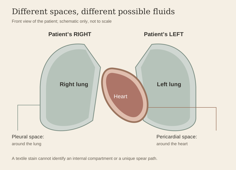
17. Where could blood and clear fluid have come from?
Pleural fluid with blood. One model proposes fluid around a lung together with blood released from a penetrated cardiac chamber or injured thoracic vessels. Zugibe favoured a pleural-fluid/right-atrial explanation. It has an intelligible anatomical basis, but the presence, size, and origin of a substantial effusion in Jesus remain inferred. Trauma does not guarantee that a visible volume of clear fluid will accumulate within the available time.3
Pericardial fluid with cardiac blood. Barbet connected the clear component to an effusion around the heart and the blood to a cardiac source. This differs from saying the person died of cardiac rupture. Fluid within the pericardial sac could be proposed without a spontaneous rupture, while a haemopericardium hypothesis introduces blood into that compartment before the spear. These accounts should not be blended into one undifferentiated “heart fluid” explanation.2, 3
Blood and separated fluid in a chest collection. A haemothorax or blood-containing effusion might present components of different appearance under some conditions. Clotting, sedimentation, fibrinolysis, dilution, position, and elapsed time would affect what drained. “Blood and water” does not establish that complete gravitational layering occurred, that blood necessarily emerged first from a lower layer, or that separation fixes the time since death.3, 7
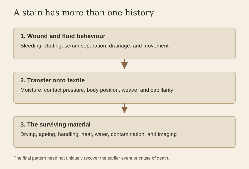
Clot-associated serum and subsequent transfer. Serum can separate during clot retraction, and a textile can receive cellular-rich and relatively clear material differently. Such a pattern concerns the formation of a stain; it does not require the visible fluids to have come from two internal cavities. This is an especially important alternative when moving from John’s reported observation to the side-region appearance on linen.7, 21, 22
The existence of several plausible sources is the central finding. A clear component is medically possible, but identifying it requires evidence beyond colour and location. No valid anatomical conclusion follows from treating the word “water” as a unique label for one body fluid.
18. Bleeding after death is possible
Blood does not instantly become immobile when the heart stops. Blood may remain fluid or later become more fluid as fibrin is broken down. Wounds can drain under gravity, compression, repositioning, or handling. Zugibe discusses postmortem oozing from injured bodies and uses it to challenge the mistaken claim that blood flow after spearing would prove the victim was alive.3
There is nevertheless an important logical limit. Passive flow is compatible with death, but a stain without visible spray is not proof of death. Bleeding during life can be slow, venous, low-pressure, or restricted by the wound. Fabric may never intercept arterial spatter produced elsewhere. Transfer to cloth may occur after a flow has already formed on skin. The absence of a particular pattern on the cloth cannot substitute for an examination of circulation.7, 10
Postmortem fluidity is also not specific for asphyxia. Nor does fibrinolysis automatically distinguish antemortem from postmortem deposition in old linen. A clot could form during life, be disturbed during removal from the cross, release fluid after death, and transfer to cloth later. Several stages may contribute to a single final mark. That sequence is plausible and experimentally examinable; its plausibility is not proof that it occurred on the Shroud.7
It follows that neither “blood flowed, therefore he was alive” nor “the flow was passive, therefore this exact death mechanism is proven” is a reliable inference. The physiology permits a range of behaviours, and the historical sequence must be assessed separately.
19. The blood evidence: chemistry, fluorescence, and interpretation
Heller and Adler reported spectroscopic and chemical findings they identified as blood in wound-associated material. Miller and Pellicori documented ultraviolet fluorescence behaviour, including features relevant to pale margins near some reddish marks. These are different kinds of evidence: chemical reactions concern sampled substances, whereas fluorescence photographs locate optical patterns on the cloth. Neither should be replaced by visual impressions alone.23, 24
The defensible formulation is that blood-associated components have been reported in examined wound-region samples. It is a further step to establish uncontaminated human origin, an individual blood group, the person’s identity, or a precise terminal condition. The supplied recent blood-transfer review explicitly rejects categorical claims that the existing record conclusively establishes human, male, AB, or diagnostically polytraumatic blood. This chapter follows that distinction.7
A fluorescent margin can be compatible with separated serum, but fluorescence alone is not chemically unique to serum. Kearse’s experiments demonstrate that illumination near 365 nm can reveal serum and plasma and can distinguish some transferred clot-associated patterns from direct application in the tested conditions. This supports experimental investigation of the Shroud margins; it does not identify the source of every margin or establish a cause of death.21
Ageing, heat, water exposure, contamination, textile structure, and sampling location matter. A serum-like ring cannot be read as a preserved laboratory result for coagulation time, bilirubin concentration, terminal acidosis, or time since death. Those would require validated measurements and controls beyond the available observations.7, 17
20. Experimental stains and competing sequences
König and colleagues examined possible formation sequences for the head stains, the lateral chest stain, and the dorsal “belt of blood.” Their experiments produced a side-stain margin using sequential application of clear biological fluid and whole blood, and a belt-like transfer through repositioning a body model. The publication appeared online in 2023 and in the 2024 issue of International Journal of Legal Medicine.22
These findings weaken any claim that the gross appearance must arise from one unique event. They do not demonstrate that the Shroud was washed, that a particular burial procedure occurred, or that the cloth was manufactured. Experimental resemblance establishes a feasible route under specified conditions. Its historical relevance depends on materials, geometry, fluid properties, contact, and comparison with alternatives.
Kearse’s humidity study adds another relevant possibility: dried blood can regain transferability in high humidity under experimental conditions. That bears on how earlier wounds might leave later marks. It does not establish the temperature or relative humidity of Jesus’s tomb, the duration of contact, or the preservation of every stain.25
The author’s recent review integrates antemortem clotting, serum separation, wound disturbance, postmortem drainage, and eventual contact with linen into a constrained model. Its value is that the stages can be tested against alternatives. Its status remains a synthesis, and later careful qualifications should take priority over stronger, less supported assertions in earlier collection papers.7, 17
21. Rigor, abdominal contour, and the body after death
The Shroud’s body position has been interpreted as compatible with rigor mortis: partially flexed knees, a forward-inclined head, and a body that may have retained aspects of its former posture. Such interpretations concern morphology after death. They cannot identify the last breath or prove that the chest was frozen at maximal inspiration. Primary relaxation, the development of rigor, repositioning, gravity, and cloth tension all intervene between suspension and the final image.2, 3, 4, 26
Chapter 13 separately examines the lower-abdominal protrusion. Its proposed relation to diaphragm-dominant breathing is relevant here only as a possible mechanical association. A contour compatible with altered thoracoabdominal mechanics is not a diagnostic sign of fatal asphyxia. The transition from active breathing on a cross to a supine body beneath linen is an unresolved part of the model.27
A three-dimensional-looking rendering does not solve that problem. Converting image intensity into height creates a relief based on assumptions about the image signal. It is not an internal scan and cannot demonstrate lung inflation, organ enlargement, cardiac rupture, or fluid within a cavity. Similarly, a seemingly composed facial expression on a processed image cannot distinguish a particular terminal disease or quantify the suffering before death.26, 27
Claims that the body left the cloth within a specific number of hours because decomposition is not apparent require independent validation. The failure to identify obvious decomposition marks is an observation about the surviving image; it is not a clock with a fixed start, known conditions, and an established detection threshold. The medical question of death should not be made to depend on an unmeasured separation interval.
22. Comparing the main mechanisms
| Proposed mechanism | Why it is considered | What is missing in this case |
|---|---|---|
| Blood/fluid loss with traumatic shock | Reported scourging and painful prolonged execution provide plausible pathways to impaired perfusion. | Measured losses, circulation, organ perfusion, and injury depth. |
| Exhaustion or posture-related respiratory failure | Injury, effort, and restraint can affect ventilation; Barbet and the JAMA reconstruction give this a central role. | A validated compulsory breathing cycle and case-specific respiratory measurements; volunteer results challenge strong versions. |
| Combined circulatory and respiratory failure | Explains interaction among several plausible stresses without requiring a single isolated lesion. | The severity and contribution of each component; the exact terminal sequence. |
| Trauma-induced coagulopathy | Recognized after severe injury and shock; proposed by Bergeron. | Coagulation testing or independently diagnostic material from Jesus. |
| Cardiac rupture/tamponade | Could produce abrupt collapse and a source of blood within the pericardium. | An infarct or other myocardial lesion, rupture, and an established pericardial collection. |
| Stress cardiomyopathy | Extreme stress offers a plausible trigger for a recognized cardiac syndrome. | Cardiac imaging, biomarkers, ECG, and exclusion of alternatives. |
| Pulmonary embolism | Trauma and immobilization motivate a vascular hypothesis. | Embolus, source thrombus, timing, or individual thrombophilia evidence. |
| Fatal spear thrust | A deep penetrating chest wound can kill a living person. | Consistency with John’s stated sequence, in which death preceded the wound. |
The table compares explanatory requirements rather than declaring a winner by vote. A simpler model is not automatically true, and a more elaborate one is not stronger merely because it contains more modern diagnoses. The appropriate question is whether a proposed addition is independently supported and whether it explains something the available evidence actually establishes.2, 3, 8, 11, 12, 14, 15, 19, 20
23. What further research could clarify
Ethical studies cannot reproduce an execution, but they can isolate parts of the problem. Supervised posture experiments can compare arm angles, partial foot loading, chest-wall movement, respiratory muscle recruitment, circulation, discomfort, and the ability to change position. Results should be reported for the tested configuration and participant population. A finding in one safe model should not be advertised as a complete simulation of crucifixion.
For the side wound, anatomical modelling can compare explicitly stated trajectories without claiming that a preferred trajectory was historically used. Blood-transfer experiments can vary fluid composition, clot age, textile structure, contact pressure, moisture, body position, and movement. Clear-fluid controls are necessary if a serum-like margin is the proposed discriminator. Heat, water exposure, and ageing controls matter when applying modern experiments to an old textile.7, 22
For the cloth itself, improved photographic documentation and spatially resolved analysis could better distinguish an optical margin from a chemically identified fluid component. Sampling would require conservation approval and a documented provenance. Interpretation should be blinded where feasible, and rival formation sequences should be tested against the same predefined features.
Even successful new work would have a limited remit. It could strengthen or weaken proposed mechanisms and establish which features are reproducible. It could not recover a missing electrocardiogram or convert an ancient narrative into a modern hospital chart. That limit makes careful experiments more valuable, because they can replace specific assumptions with measured constraints.
24. The strongest forensic synthesis
The historical question and the physiological question end at different levels of resolution. The Gospel accounts present Jesus as dead before burial, with Mark describing official confirmation and John describing a subsequent side-piercing episode. Medical reconstruction asks how the reported violence could have brought about that death. The records allow credible mechanisms, but not a uniquely demonstrated clinical diagnosis.1
The strongest broad framework is a body subjected to interacting stresses. Blood and fluid loss could reduce circulation. Pain and sustained effort could increase metabolic demand. Posture, exhaustion, and possible thoracic injury could impair respiratory effectiveness. Circulatory failure and respiratory compromise could then aggravate each other, with death occurring when compensation failed. This is the synthesis adopted here as a plausible explanatory framework, not a claim of established medical consensus or a measured sequence.
Specific alternatives remain open. Cardiac rupture, stress cardiomyopathy, pulmonary embolism, and coagulation disturbance require findings that are unavailable. Asphyxia remains a prominent historical explanation, but the claim that a compulsory raising-and-sagging cycle is proven must confront experimental and historical objections. Shock is physiologically plausible without supplying an invented blood-loss total.
The side wound gives a separate problem: how to interpret a reported release of blood and clear fluid and a textile stain with differing components. Several anatomical and transfer mechanisms can account for aspects of those observations. They do not uniquely diagnose the terminal event, establish an exact time since death, or authenticate the Shroud by themselves.
For a Christian reader, the medical inquiry can make the bodily reality of crucifixion more intelligible without reducing the meaning of Jesus’s death to a physiological formula. Theological claims about voluntary self-giving belong to the interpretation of the event. The forensic task is narrower: to explain what the evidence supports, identify what remains hypothetical, and let neither certainty of faith nor skepticism substitute for an unperformed examination.
Source and illustration notes
The supplied local article collection was inventoried and screened for relevance. The medical, blood-transfer, postmortem, image-interpretation, and draping papers were considered alongside the relevant sections of the supplied books. Duplicate files and unrelated papers do not constitute additional evidence. Barbet’s and Zugibe’s original books were consulted directly; Malantrucco’s mechanism is discussed through the supplied Forensic Summary, rather than represented as a directly verified reading of his complete book. This is an extensive critical narrative synthesis, not a systematic review or a claim that every book in the broader library was read cover to cover.
Figures 1–2 reuse author-supplied Atlas/book assets. Figures 3–5 are original explanatory diagrams for this chapter. Reconstructions are identified as interpretations; no generated scene is presented as a historical photograph or experimental observation. Artificial intelligence assisted research organization, drafting, and diagram preparation. The chapter requires the author’s final scholarly review.
References and further reading
References are numbered in order of first appearance. Source type and inferential limits are supplied to make the evidence traceable.
- Historical sourceThe canonical Gospels: Matthew 27; Mark 15, particularly 15:25, 33–37, 42–45; Luke 22:39–46 and 23; John 19:1–42. Read source ↗.
Primary narratives; their time statements and distinct emphases are not treated as a synchronized clinical record. ↩
- Book consultedBarbet P. A Doctor at Calvary: The Passion of Our Lord Jesus Christ as Described by a Surgeon. Translated by the Earl of Wicklow. English edition, 1953; subsequent reprints. Chapters on the cause of death and the side wound, and Appendix II by P. J. Smith.
Original supplied PDF consulted, including the dissenting opinion in Appendix II. Claims are attributed to Barbet rather than treated as established findings. ↩
- Books and investigator accountZugibe FT. The Crucifixion of Jesus: A Forensic Inquiry. M. Evans, 2005, especially the chapters on asphyxiation, the cause of death, and blood and water. Also The Cross and the Shroud, revised edition, Paragon House, 1988; and “Pierre Barbet Revisited,” Sindon, new series, no. 8, 1995. Read source ↗.
Both supplied books were consulted. The linked 1995 account provides an accessible statement of Zugibe’s challenge. Related study: “Death by Crucifixion,” Canadian Society of Forensic Science Journal 17(1), 1–13 (1984), doi:10.1080/00085030.1984.10757355. ↩
- Author-supplied bookGrasso O. The Shroud of Turin: A Forensic Summary of the Evidence. Third edition, supplied PDFs 3 Forensic Summary 3nd_ed and Forensic_Summary_3nd_ed. Read source ↗.
Relevant sections include blood evidence, crucifixion anatomy, “The Final Cascade” (PDF pp. 228–238), and deposition/burial. These editions are related authorial syntheses, not independent experiments. ↩
- Author-supplied bookGrasso O. The Image That Should Not Exist: A Scientific Investigation of the Shroud of Turin. Supplied edition.
Medical-examination and crucifixion discussion, particularly PDF pp. 202–212. Unverified precision and claims about consensus were not carried forward as established facts. ↩
- Author-supplied bookGrasso O. From Forensics to Faith. 2026 supplied edition.
Relevant blood, medical, and crucifixion sections were consulted. Repetition across the author’s books is not counted as independent corroboration. ↩
- Author’s critical reviewGrasso O. Blood Transfer, Serum Halos, and Postmortem Flow on the Shroud of Turin: A Critical Forensic and Hematological Narrative Review. Supplied manuscript. Read source ↗.
Integrates clotting, transfer, rehydration, postmortem fluidity, and competing models; explicitly distinguishes plausible sequences from unique proof. ↩
- Critical medical reviewMaslen MW, Mitchell PD. Medical theories on the cause of death in crucifixion. Journal of the Royal Society of Medicine. 2006;99(4):185–188. Read source ↗.
Evaluates competing explanations and limits of historical, archaeological, and experimental evidence. Open text: PMC1420788. ↩
- Historical sourceJosephus. Life 420–421. Translation by William Whiston. Read source ↗.
Three crucified acquaintances were removed and treated; two died and one recovered. Their suspension duration and injury burden are unspecified. ↩
- Shroud image interpretationBucklin R. An autopsy on the man of the Shroud. 1997. Read source ↗.
The linked presentation is a simulated forensic examination of the image, not a literal autopsy. Its interpretations are attributed to the author. ↩
- Medical reconstructionEdwards WD, Gabel WJ, Hosmer FE. On the physical death of Jesus Christ. JAMA. 1986;255(11):1455–1463. Read source ↗.
Influential historical-medical reconstruction proposing hypovolaemic shock and exhaustion asphyxia; not an examination of the body. ↩
- Medical hypothesis reviewBergeron JW. The crucifixion of Jesus: review of hypothesized mechanisms of death and implications of shock and trauma-induced coagulopathy. Journal of Forensic and Legal Medicine. 2012;19(3):113–116. Read source ↗.
Proposes shock and coagulopathy as possible mechanisms while acknowledging that the precise cause cannot be proven. ↩
- Physiological experimentRauch S, Schenk K, Strapazzon G, et al. Suspension syndrome: a potentially fatal vagally mediated circulatory collapse—an experimental randomized crossover trial. European Journal of Applied Physiology. 2019;119:1353–1365. Read source ↗.
Healthy harness-suspension study; used only as a limited analogy. A correction was published: doi:10.1007/s00421-019-04141-6. ↩
- Critical medical reviewMcGovern TW, Kaminskas DA, Fernandes ES. Did Jesus die by suffocation? An appraisal of the evidence. The Linacre Quarterly. 2023;90(1):64–79; online 2022. Read source ↗.
Challenges the suffocation explanation and the use of suspension-torture analogies. Open text: PMC10009142. ↩
- Survey of medical viewsHabermas G, Kopel J, Shaw BCF. Medical views on the death by crucifixion of Jesus Christ. Proceedings (Baylor University Medical Center). 2021;34(6):748–752. Read source ↗.
doi:10.1080/08998280.2021.1951096. The authors’ assessment of the prevalence of asphyxia views is distinguished from experimental proof. ↩
- General trauma physiologyMoore EE, Moore HB, Kornblith LZ, et al. Trauma-induced coagulopathy. Nature Reviews Disease Primers. 2021;7:30. Read source ↗.
Modern general physiology, not a diagnosis of Jesus. Correction: doi:10.1038/s41572-022-00360-y. ↩
- Author’s earlier analysisGrasso O. Pre-Mortem and Post-Mortem Blood on the Shroud of Turin: A Convergent Constraint Analysis. 25 January 2026. Read source ↗.
Read alongside the later, more qualified blood-transfer review. Strong species-identification and diagnostic claims are not adopted uncritically. ↩
- Indirect book citationMalantrucco L. Il silenzio della Sindone: analisi della morte di un uomo chiamato Gesù. 2010.
The haemopericardium proposal is discussed here through Grasso’s Forensic Summary, chapter “The Final Cascade.” Malantrucco’s complete book was not directly consulted for this chapter. ↩
- Recent medical hypothesisMangione S. Did Jesus die of Takotsubo cardiomyopathy? A medical revisiting of the Gospels. Journal of General Internal Medicine. 2024;39:3342–3343. Read source ↗.
PMID:39299976. A retrospective hypothesis. Subsequent commentary and reply: doi:10.1007/s11606-024-09307-3 and doi:10.1007/s11606-024-09304-6 (2025). ↩
- Medical hypothesisBrenner B. Did Jesus Christ die of pulmonary embolism? Journal of Thrombosis and Haemostasis. 2005;3(9):2130–2131. Read source ↗.
A proposed vascular explanation, not case-specific evidence of thrombosis or a genetic finding. ↩
- Laboratory experimentKearse KP. Ultraviolet 365 as an alternative light source for detection of blood serum. Journal of Forensic Sciences. 2020;65(5):1716–1721. Read source ↗.
Tests of serum/plasma detection and clot-associated transfer; not a new test of the Shroud. ↩
- Shroud-related experimentsKönig L, Schmölders R, Jühling M, Reckert A, Heger A, Ritz-Timme S. Some experiments and remarks regarding the possible formation of blood stains on the Turin Shroud: stains attributed to the crown of thorns, the lance wound and the belt of blood. International Journal of Legal Medicine. 2024;138:229–238; online 2023. Read source ↗.
Experimental alternatives for stain formation. Reproduction of a pattern does not establish that it occurred historically. Open text: PMC10772002. ↩
- Sample analysisHeller JH, Adler AD. Blood on the Shroud of Turin. Applied Optics. 1980;19(16):2742–2744. Read source ↗.
Chemical and spectroscopic analysis of sampled wound-associated material. Identification, provenance, species specificity, and the cause of death are separate questions. ↩
- Optical documentationMiller VD, Pellicori SF. Ultraviolet fluorescence photography of the Shroud of Turin. Journal of Biological Photography. 1981;49(3):71–85. Read source ↗.
Original UV photographic documentation; fluorescence patterns are not equivalent to spatially specific biochemical identification. ↩
- Laboratory experimentKearse KP. The effect of humidity on blood serum pattern formation and blood transfer. Journal of Forensic Science and Research. 2023;7:040–048. Read source ↗.
Experiments on drying and rehydration. Conditions are not measurements of Jesus’s tomb. ↩
- Author’s draping analysisGrasso O. The Draping Configuration of the Shroud of Turin During Image Formation: A Synthesis of Geometric, Photochemical, and Radiation Hypotheses. 18 September 2025. Read source ↗.
Used for cloth/body geometry and transfer constraints, not as independent proof of a particular burial procedure. ↩
- Author’s critical reviewGrasso O. Lower-Abdominal Protrusion in Crucifixion and the Turin Shroud: A Critical Review of Thoracoabdominal Mechanics, Suspension Physiology, Postmortem Change, and Three-Dimensional Image Interpretation. 8 August 2026. Read source ↗.
Full discussion remains in Chapter 13. A compatible abdominal contour is not a specific diagnosis of fatal asphyxia. ↩